Introduction:
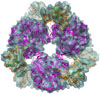
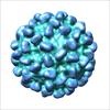
Cryo-electron microscopy (Cryo-EM) and three-dimensional (3D) reconstruction techniques are used in our research to study the architecture of large macromolecular assemblies of three types: (i) multi-enzymatic complexes, (ii) viruses and capside proteins, and (iii) composite nano-particles. Several technological advances led to a recent explosive growth in this field and have allowed researchers to study molecular structures and interactions at sub-nanometer resolutions. Therefore, 3D Cryo-EM approach combined with other data from structural biology techniques (Light diffusion, SAXS, SANS, X-Ray crystallography, RMN, molecular modeling, normal mode analysis) are now used to solve protein structure, and to provide a dynamic multiscale vision of large macromolecular assemblies in action.
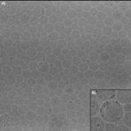
The most important input from 3D Cryo-EM, is that it provides a global sub-nanometric map of biological samples preserved in a near native state. For this, samples are flash frozen in their physiological environmental conditions (hydrated, pH, salts, detergents, cofactors, ATP analogs, RNA, DNA, etc
) by rapid plunging into liquid ethane. In such condition ice crystals formation damaging for the sample is prevented, and the sample is just fixed in a vitreous ice preserving medium. Specimen grids are then maintained at liquid nitrogen temperature, while inserted and observed in the electron microscope, and no stain or chemical fixation is used during this whole process. We now have access to two high resolution JEOL 2100 and 2100F microscopes, equipped with 4K and 2K Gatan Ultrascan CCD cameras used for automated computer controlled data collection, and we also use Kodak So163 photographic films for the highest resolution data. Depending on the structural heterogeneity of samples, sets of 10.000 to 200.000 individual images may be used to get a 3D map at sub-nanometer resolution.
Image processing and fast parallel computing techniques are continuously being developed to extract and process the signal from noisy image data sets. For example, (i) contrast variations resulting from Contrast Transfer Function (CTF) of electron microscopes must be accurately estimated and corrected; (ii) directions of projection of large sets of noisy images must be accurately refined by iterative alignments; (iii) and once a 3D Cryo-EM map is obtained, this information needs to be combined with other data (X-ray, NMR, NMA, etc..) to by related with motion and activity of these large complexes. Finally, these data must be deposited for the benefit of all research communities in the Macromolecular Structural Database (MSD).
Our current research efforts are focused on:
- The two multi-enzymatic complexes (Glutamate synthase of bacteria responsible for their ammonium metabolism, and the mammalian phosphorylase kinase, a key enzyme in glycogen metabolism and a potential target for new generations of anti-diabetic drugs.
- Newly discovered viruses infecting hyperthermophile and hallophile archaebacteria.
- Calcium and pH dependant swelling mechanism of plant viruses.
- The observation of quantum dots and new composite nanoparticles using cryoEM techniques.
- Fast and accurate CTF estimation and correction.
- Development of image processing algorithms for 3D high-resolution reconstruction (e.g, estimation and correction of the CTF, computation of 3D structures in presence of structural heterogeneity).
 |
Jonic S, Sorzano COS, Cottevieille M, Larquet E, Boisset N. A novel method for improvement of visualization of power spectra for sorting cryo-electron micrographs and their local areas, J Struct Biol , vol. 157, pp. 156167 , 2007. |
We also develop technical project on: - Computer-controlled flash freezing of samples
- Fast digitization of micrographs and automated data collection.
CMET team |
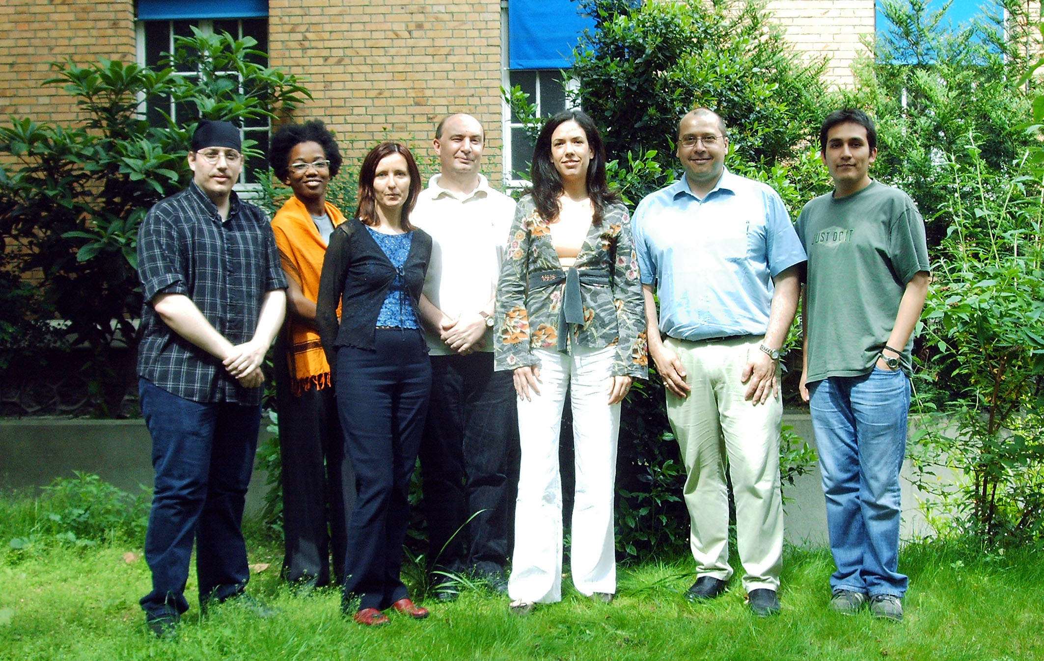 |
Left to right: Thierry, Cathelène, Slavica, Eric, Magali, Nicolas, Ricardo |
|